

1. Definition
Proximal Femur Nailing (PFN) and Tibia and Leg Bone Nailing are orthopedic procedures used to:
2. Purpose
3. When is PFN or Tibia Nailing Recommended?

Used for hip and upper femur fractures.
Intramedullary nail inserted into the femoral canal.
Provides stability and alignment.
Common for elderly patients with hip fractures.
Treats tibia shaft fractures.
Intramedullary nails inserted into the tibia bone.
Stabilizes the leg and prevents misalignment.
Less common than tibia nailing.
Used for complex or multi-fragment fractures.
Provides stability alongside tibia fixation.
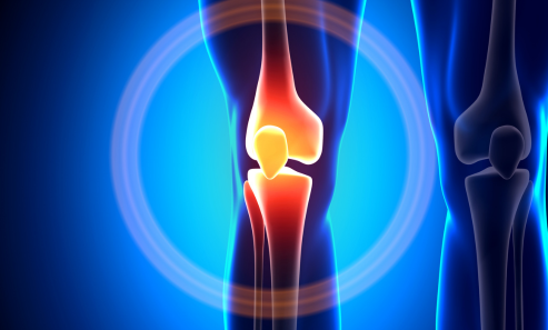
For distal femur fractures (near the knee).
Nail inserted through the knee joint.
Stabilizes the femur and allows early mobility.
Combination of external fixation and IM nailing.
Used in severe open fractures.
Prevents infection and enhances stability.
Intense pain in the thigh, leg, or knee.
Swelling and tenderness around the fracture site.
Visible misalignment or deformity.
Affected leg may appear shorter or bent.
Difficulty standing or walking.
Loss of mobility or function.
Internal bleeding may cause bruising.
Skin may appear purple or red.
Bone fragments protruding in open fractures.
Limited range of motion.
Instability in the affected leg.
Calcium and Vitamin D-rich diet.
Reduces the risk of osteoporosis.
Enhances bone density and muscle support.
Reduces fall risks.
Use handrails, non-slip mats, and proper lighting.
Especially important for the elderly.
Bone density screening for at-risk individuals.
Osteoporosis medications.
Use knee pads, helmets, and guards during sports.
Reduces fracture risks.
Surgical realignment of fractured bones.
Prevents deformity and promotes healing.
Intramedullary nails inserted into bone canals.
Provides stability and support.
For severe fractures with bone loss.
Graft promotes bone regeneration.
Imaging tests: X-rays, CT scans, or MRI.
Anesthesia consultation: General or spinal anesthesia.
Fasting before surgery.
Incision made over the fracture site.
Fracture exposed and realigned.
Intramedullary nail inserted into bone canal.
Screws or bolts secure the nail.
Incision closed with stitches or staples.
Bandages and dressings applied.
1-3 days of hospitalization.
Pain management with analgesics.
Partial weight-bearing with crutches.
Gradual transition to full weight-bearing.
Antibiotics to prevent infection.
Blood thinners to prevent clots.
Regular X-rays to monitor bone healing.
Physical therapy for mobility restoration.
Standard procedure for tibia, femur, or leg bone fractures.
Nails and screws secure the bone.
For distal femur fractures.
Nail inserted through the knee joint.
Used in open fractures.
Temporary fixation with external frame.


