ORIF (Open Reduction Internal Fixation)

img
img

What is ORIF (Open Reduction Internal Fixation)?

img
Definition

Open Reduction Internal Fixation (ORIF) is a surgical procedure used to:

  • Repair and stabilize broken bones.
  • Involves making an open incision to align the fractured bones (open reduction).
  • Uses metal plates, screws, rods, or pins to hold the bones in place (internal fixation).
Purpose
  • Restore bone alignment and promote healing.
  • Prevent deformity or loss of function.
  • Used for complex or severe fractures that cannot be treated with casting alone.
When is ORIF recommended?
  • Severe or displaced fractures.
  • Fractures with joint involvement.
  • Multiple bone fragments (comminuted fracture).
  • Failure of non-surgical treatments (e.g., splints, casts).
  • Fractures that affect mobility or stability.

Types of ORIF Procedures

img
ORIF for Long Bones

Femur, tibia, humerus, radius, ulna, or fibula fractures.

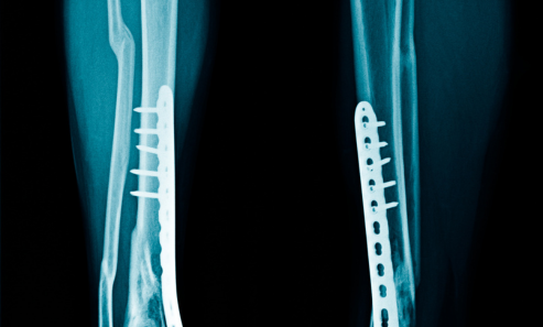
Plates, screws, or rods inserted along the bone.

Restores proper alignment and function.

img
ORIF for Joint Fractures

Ankle, wrist, elbow, shoulder, or knee fractures.

Fixation devices stabilize the joint.

Prevents arthritis or stiffness.

img
ORIF for Hip Fractures

Hip fractures in elderly patients.

Screws, plates, or rods inserted.

Prevents hip displacement and promotes mobility.

img
ORIF for Pelvic Fractures

Complex pelvic fractures require internal fixation.

Reduces pain and prevents complications.

Plates and screws stabilize the pelvis.

img
ORIF for Hand and Foot Fractures

Used for metacarpal or phalangeal fractures.

Helps restore fine motor functions.

Plates and screws secure small bones.

Symptoms Indicating the Need for ORIF

Doctors may recommend angiography if a patient experiences:

img

Severe Pain and Swelling

  • Persistent and intense pain around the fracture.
  • Swelling, tenderness, and redness.
img

Bone Deformity

  • Misaligned or visibly distorted bones.
  • Uneven limb length.
img

Loss of Mobility or Function

  • Inability to move the affected area.
  • Joint stiffness or loss of range of motion.
img

Open Fractures

  • Bone pierces the skin.
  • Risk of infection and tissue damage.
img

Non-Healing Fractures

Risk Factors for ORIF

img

Causes Leading to ORIF

Traumatic Injuries
  • Car accidents or falls.
  • Cause complex or displaced fractures.
Sports-Related Fractures
  • High-impact sports (football, skiing).
  • Lead to joint or bone fractures.
Osteoporosis
  • Weakens bones in elderly individuals.
  • Increases risk of hip and wrist fractures.
Workplace or Industrial Accidents
  • Severe fractures from machinery accidents.
  • Requires ORIF for stabilization.
Pathological Fractures
  • Bone tumors or cancer.
  • Weaken bones and cause fractures.

How to Prevent the Need for ORIF?

img
Bone Health Maintenance

Calcium and Vitamin D-rich diet.

Strengthens bones and prevents fractures.

img
Regular Exercise

Weight-bearing exercises enhance bone density.

Improves balance and reduces fall risk.

img
Fall Prevention

Use handrails, non-slip mats, and proper lighting.

Especially important for the elderly.

img
Treat Osteoporosis

Bone density screening for at-risk individuals.

Use of osteoporosis medications.

img
Protective Gear

Wear helmets and padding during high-risk activities.

Reduces fracture risks.

Treatment Through ORIF

img
Bone Realignment

Surgical realignment of fractured bones.

Reduces deformity and restores function.

img
Internal Fixation

Plates, screws, rods, or wires inserted.

Provides stability during healing.

img
Bone Grafting

Used for severe fractures or bone loss.

Bone graft enhances healing.

ORIF Procedure Steps

img
Pre-Procedure Preparation

Imaging tests: X-rays, CT scans, or MRI.

Anesthesia consultation: General or regional anesthesia.

Fasting before surgery.

img
During the Procedure
Steps 1:

Incision made over the fracture site.

Bone exposed for realignment.

Steps 2:

Fractured bones are realigned.

Internal fixation with screws, plates, rods, or wires.

Steps 3:

Incision closed with stitches or staples.

Bandages and dressings applied.

img
Hospital Stay

1-3 days of hospitalization.

Pain management with analgesics.

Restricted Mobility

Use of crutches or walkers for lower limb fractures.

Limited weight-bearing for several weeks.

Medications

Antibiotics to prevent infection.

Blood thinners to prevent clots.

Follow-Up Care

Regular X-rays to monitor bone healing.

Physical therapy for mobility restoration.

img
ORIF with Plates and Screws

Plates and screws hold bones in place.

Common for long bone fractures.

ORIF with Intramedullary Rods

Rod inserted inside the bone canal.

Stabilizes femur, tibia, or humerus fractures.

ORIF with Wires or Pins

Used for small bone fractures.

Common in hand or foot fractures.