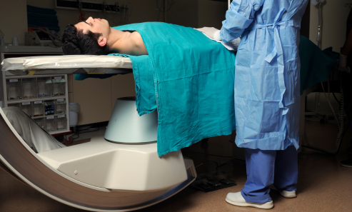


Angiography is a medical imaging test used to examine blood vessels (arteries and veins) for blockages, narrowing, or abnormalities. A contrast dye is injected into the bloodstream, and X-rays, CT scans, or MRIs capture detailed images of blood flow. It helps diagnose conditions like heart disease, stroke, aneurysms, and peripheral artery disease.

Examines heart arteries to detect blockages.

Checks blood vessels in the brain.

Used to detect blood clots in the lungs.

Examines arteries in the legs, arms, or other areas.

Evaluates blood flow in the kidneys.

Evaluates blood flow in the kidneys.

Used to examine blood flow in the retina (eye).

Assesses blood vessels supplying the intestines.
Chest pain (angina)
Shortness of breath
Frequent dizziness or fainting

Numbness or weakness in limbs
Sudden vision problems or slurred speech
Unexplained pain in arms, legs, or abdomen
High blood pressure affecting the kidneys
While generally safe, angiography carries some risks, including:

Angiography is performed when conditions like these are suspected:
While some conditions may be unavoidable, you can lower the risk by:
If angiography reveals blockages, possible treatments include:
Medications – Blood thinners, cholesterol-lowering drugs, or blood pressure medications.
Lifestyle changes – Improved diet, exercise, and stress management.
Angioplasty and Stent Placement – A procedure to open narrowed arteries.
Bypass Surgery – Creating a new route for blood flow around blocked arteries.
Thrombectomy – Removing a blood clot causing blockage.

Angiography can be performed using different methods:

A catheter is inserted into an artery to inject contrast dye and take X-ray images.

Uses a CT scan to get detailed 3D images of blood vessels.

Uses MRI technology for a detailed look at blood vessels without X-ray radiation.